Arteries Diagram : Brachial Artery Anatomy Britannica : Blood carried by arteries is usually highly oxygenated, having just left the lungs on its way to the body's tissues.
byAdmin-
0
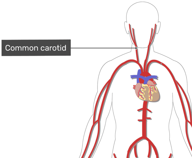
Arteries Diagram : Brachial Artery Anatomy Britannica : Blood carried by arteries is usually highly oxygenated, having just left the lungs on its way to the body's tissues.. Arteries and veins are two of the body's main type of blood vessels. Plaques are clumps of cholesterol, calcium, fibrous tissue and other cellular debris that gather at microscopic injury sites within the artery. Coronary arteries supply oxygenated blood to the heart muscle. There are three arteries of the heart, including pulmonary artery, aorta, and coronary arteries. From this trunk, several vessels arise, which go on to supply the neck.
This process is called atherosclerosis. In this image, you will find right gastric artery, common hepatic artery, celiac trunk, left gastric artery, splenic artery, splenic vein, pancreas, suprarenal vein, renal vein, renal artery, inferior mesenteric vein , gonadal vein, gonadal artery, two alternative position of artery, left colic artery. Learn vocabulary, terms, and more with flashcards, games, and other study tools. Daniel nelson on january 1, 2019 1. The abdominal aorta bifurcates at the level of the fourth lumbar vertebra to form the two common iliac arteries, each of which further branches into the external and the internal iliac artery.
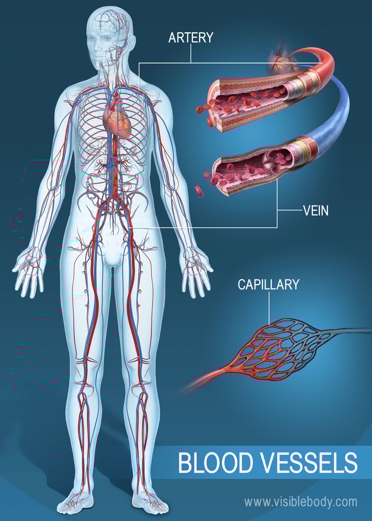
Blood Vessels Circulatory Anatomy from www.visiblebody.com Simplified diagram of the human arterial system in anterior view. As with veins, arteries are comprised of three layers: An artery (plural arteries) (from greek ἀρτηρία (artēríā) 'windpipe, artery') is a blood vessel that takes blood away from the heart to one or more parts of the body (tissues, lungs, brain etc.). Other arteries of the neck. After receiving blood directly from the left ventricle of the heart, the. It can also help them in getting an overview of artery vs. Like maps, the various diagrams emphasize different aspects. There are three arteries of the heart, including pulmonary artery, aorta, and coronary arteries.
In the organs both arteries and veins divide to form arterioles and venules respectively.
Human body artery diagram in detail. The heart consists of a range of tissues. Check out our heart diagrams, quizzes and worksheets. Some are more conceptual, others focus on branching, while still others attempt to preserve a spatial representation. Blood is transported in arteries, veins and capillaries. In this image, you will find right gastric artery, common hepatic artery, celiac trunk, left gastric artery, splenic artery, splenic vein, pancreas, suprarenal vein, renal vein, renal artery, inferior mesenteric vein , gonadal vein, gonadal artery, two alternative position of artery, left colic artery. Because the rest of the body, and most especially the brain, needs a steady supply of oxygenated blood that is free of all but the slightest. The abdominal aorta bifurcates at the level of the fourth lumbar vertebra to form the two common iliac arteries, each of which further branches into the external and the internal iliac artery. Abdomen arteries, veins, and duct diagram. Coronary arteries supply blood to the heart muscle. These vessels are channels that distribute blood to the body. Blood is pumped from the heart in the arteries. This process is called atherosclerosis.
Plaques are clumps of cholesterol, calcium, fibrous tissue and other cellular debris that gather at microscopic injury sites within the artery. It can also help them in getting an overview of artery vs. Carotid artery disease is caused by a buildup of plaques in arteries that deliver blood to your brain. You can imagine the aorta and ivc as the two trees, with all. Blood is transported in arteries, veins and capillaries.
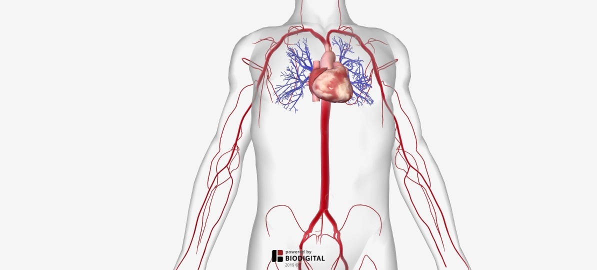
Arteries Of The Body Picture Anatomy Definition More from human.biodigital.com The neck is supplied by arteries other than the carotids. Some are more conceptual, others focus on branching, while still others attempt to preserve a spatial representation. As with veins, arteries are comprised of three layers: The two exceptions are the pulmonary and the umbilical arteries, which carry deoxygenated blood to the organs that oxygenate it (lungs and placenta. Because the rest of the body, and most especially the brain, needs a steady supply of oxygenated blood that is free of all but the slightest. John bavosi/science photo library/getty images. This is the opposite function of veins, which transport blood to the heart. In this image, you will find external carotid artery, internal carotid artery, vertebral artery, aorta and arch, pulmonary artery, cardiac artery, thoracic aorta, celiac trunk, superior mesenteric artery, renal artery, gonadal artery, inferior mesenteric artery, common iliac artery, external iliac artery.
Smartdraw includes 1000s of professional healthcare and anatomy chart templates that you can modify and make your own.
Only 3 available and it's in 2 people's carts. As with veins, arteries are comprised of three layers: Bodytomy provides a labeled iliac artery diagram to help you understand the anatomy and function of the common iliac. This process is called atherosclerosis. Veins bring blood from different body parts to the heart. Plaques are clumps of cholesterol, calcium, fibrous tissue and other cellular debris that gather at microscopic injury sites within the artery. Creating a freehand diagram of arteries and veins can be troublesome. Arteries and veins are main blood vessels. In the organs both arteries and veins divide to form arterioles and venules respectively. Arteries, veins, and the heart are the main parts of the system. Arteries of the lower limb thigh leg foot the main artery of the lower limb is femoral artery it is a continuation of the external iliac artery terminal branch of the abdominal aorta the arteries and veins of the leg smartdraw arteries and veins of the leg create healthcare diagrams like this example called arteries and veins of the leg in minutes with smartdraw. To understand the system, the students need to create their diagrams. Arteries of the head and neck diagram art print vintage anatomy art print on tea stained paper dog art dog s wfh office art.
Coronary circulation is the circulation of blood in the blood vessels that supply the heart muscle (myocardium). Cardiac veins then drain away the blood after it has been deoxygenated. In this image, you will find external carotid artery, internal carotid artery, vertebral artery, aorta and arch, pulmonary artery, cardiac artery, thoracic aorta, celiac trunk, superior mesenteric artery, renal artery, gonadal artery, inferior mesenteric artery, common iliac artery, external iliac artery. There are three arteries of the heart, including pulmonary artery, aorta, and coronary arteries. Arteries of the leg diagram.
Major Systemic Arteries from www.getbodysmart.com There are two types of them: In arteries, the tunica media, which contains smooth muscle cells and elastic tissue, is thicker than that of veins so it can modulate vessel caliber and thus control. Carotid artery disease is caused by a buildup of plaques in arteries that deliver blood to your brain. Learn vocabulary, terms, and more with flashcards, games, and other study tools. The abdominal aorta bifurcates at the level of the fourth lumbar vertebra to form the two common iliac arteries, each of which further branches into the external and the internal iliac artery. John bavosi/science photo library/getty images. You can imagine the aorta and ivc as the two trees, with all. In this image, you will find external carotid artery, internal carotid artery, vertebral artery, aorta and arch, pulmonary artery, cardiac artery, thoracic aorta, celiac trunk, superior mesenteric artery, renal artery, gonadal artery, inferior mesenteric artery, common iliac artery, external iliac artery.
Arteries and veins of the arm.
The first branch of the thyrocervical trunk is the inferior thyroid artery. This is the opposite function of veins, which transport blood to the heart. From this trunk, several vessels arise, which go on to supply the neck. Blood carried by arteries is usually highly oxygenated, having just left the lungs on its way to the body's tissues. Abdomen arteries, veins, and duct diagram. Two major coronary arteries branch off from the aorta near the point where the aorta and the left ventricle meet. An artery (plural arteries) (from greek ἀρτηρία (artēríā) 'windpipe, artery') is a blood vessel that takes blood away from the heart to one or more parts of the body (tissues, lungs, brain etc.). The capillaries connect the two types of blood. Human body artery diagram in detail. Arteries are quite tough on the outside but are smooth on the inside. In this image, you will find right gastric artery, common hepatic artery, celiac trunk, left gastric artery, splenic artery, splenic vein, pancreas, suprarenal vein, renal vein, renal artery, inferior mesenteric vein , gonadal vein, gonadal artery, two alternative position of artery, left colic artery. Arteries of the head and neck diagram art print vintage anatomy art print on tea stained paper dog art dog s wfh office art. Because the rest of the body, and most especially the brain, needs a steady supply of oxygenated blood that is free of all but the slightest.